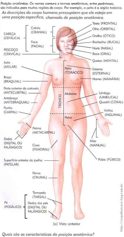
A posição anatômica é uma convenção adotada em anatomia para descrever as posições especiais dos órgãos, ossos e demais componentes do corpo humano. Na posição anatômica, o corpo estudado deve ficar ereto (de pé), calcanhares unidos, com os olhos voltados para o horizonte, os pés também apontados para frente e perpendiculares ao restante do corpo, braços estendidos e aplicados ao tronco e com as palmas das mãos voltadas para frente (os dedos estendidos e unidos).
Deve-se notar que não é a posição normal dos braços, que normalmente ficariam em torção mais ou menos medial (com as palmas voltadas para o corpo).
O corpo humano na posição anatômica pode ser dividido conceitualmente em planos.
O plano medial, sagital ou sagital medial passa através do eixo mais longo que cruza o corpo, dos pés até a cabeça; este plano separa o corpo em antímeros direito e esquerdo. O que quer que esteja situada próximo a este plano é chamado medial, e o que está longe dele, lateral.
Um plano parasagital é paralelo ao plano medial, e muitas vezes dá-se a denominação de plano sagital ao plano medial.
O plano frontal ou plano coronal passa também pelo eixo maior (dos pés à cabeça), mas é perpendicular ao plano medial, separando a frente do corpo, ou ventre, da parte de trás, ou dorso. Algo em posição à frente do plano frontal é chamadas antgerior, ao passo que algo situado atrás desse plano é chamado posterior. (veja figura 03 e figura 04)
O plano horizontal, transverso ou axial atravessa o eixo menor do corpo, do dorso até o ventre , isto é, da posição posterior para a anterior. Denomina-se em superior e em inferior. Os nomes comuns e os termos anatômicos de várias regiões do corpo estão na figura 01 (delimitações).
Também encontramos as regiões dorsal e ventral figura 02
Regiões e Quadrantes AbdominopélvicosCavidades do Corpo
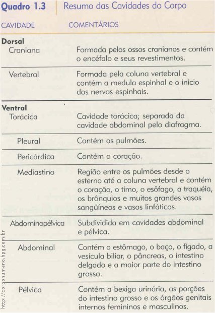
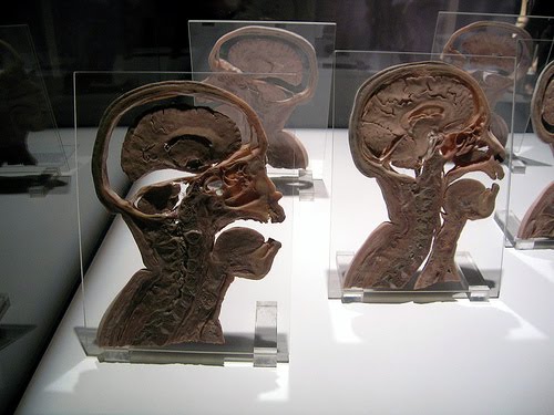
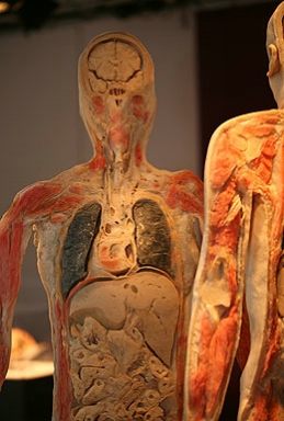
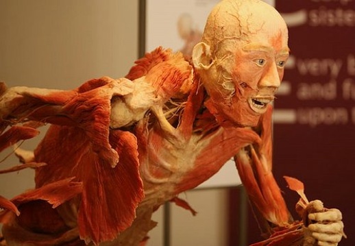
Os espaços dentro do corpo que contêm os órgãos internos são chamados de cavidades do corpo. As cavidades ajudam a proteger, isolar e sustentar os órgãos internos. A figura 05 mostra as duas principais cavidades do corpo: dorsal e ventral.
A cavidade dorsal do corpo está localizada próxima à superfície posterior ou dorsal do corpo. Ela é composta por uma cavidade craniana, que é formada pelos ossos cranianos e contém o encéfalo e suas membranas (chamadas de meninges), e por um canal vertebral que é formado pelas vértebras (ossos individuais) da coluna vertebral e contém a medula espinhal e suas membranas (também chamadas de meninges), bem como o começo (raízes) dos nervos espinhais.
A cavidade ventral do corpo está localizada na porção anterior ou ventral (frontal) do corpo e contém órgãos coletivamente chamados de vísceras. Como a cavidade dorsal, a cavidade ventral do corpo apresenta duas subdivisões principais - uma porção superior, chamada de cavidade torácica, e uma porção inferior, chamada de cavidade abdominopélvica.
O diafragma (diaphragma = partição ou parede), uma camada muscular em forma de domo e importante músculo da respiração, divide a cavidade ventral do corpo em cavidades torácica e abdominopélvica.A cavidade torácica contém duas cavidades pleurais em torno de cada pulmão, e a cavidade pericárdica (peri = em volta; cardi = coração), espaço em torno do coração (Figura 06).
O mediastino (medias = meio; stare = parar, estar), na cavidade torácica, contém uma massa de tecidos entre os pulmões que se estende do osso esterno à coluna vertebral (Figura 06). O mediastino inclui todas as estruturas na cavidade torácica, excetoos próprios pulmões.
Entre as estruturas localizadas no mediastino estão o coração, o timo, o esôfago, a traquéia e muitos grandes vasos sangUíneos, como a aorta.A cavidade abdominopélvica, como o nome sugere, está dividida em duas porções, embora nenhuma estrutura específica as separem (veja a Figura 6).
A porção superior, a cavidade abdominal, contém o estômago, o baço, o fígado, a vesícula biliar, o pâncreas, o intestino delgado e a maior parte do intestino grosso. A porção inferior, a cavidade pélvica, contém a bexiga urinária, porções do intestino grosso e os órgãos genitais internos. A cavidade pélvica está localizada entre dois planos imaginários, que estão indicados pelas linhas tracejadas da Figura 5.a.
Um resumo das cavidades do corpo é apresentado no Quadro 1.3.
Veja na figura 07: Plano Frontal, Plano Sagital, Plano Transversal, Plano Obliquo
Fonte:
http://www.sogab.com.br/floresdias/generalidadesII.htm
http://www.fernandosantiago.com.br/anato.htm
http://www.bartleby.com/107/
http://blog.uncovering.org/archives/2007/06/o_corpo_humano.html
sábado, 29 de agosto de 2009
Anatomia do Corpo Humano
Objetivo
Destinado a estudantes que necessitam de um referencial de estudo da anatomia para a sua formação. Disponibilizo os links na página para uma consulta prévia. Passe o mouse no link e clic para ser direcionado ao tema relativo à antomia humana.
É um excelente atlas que encontrei na WEB...
Vale a pena você verificar. Será uma fonte rica no que diz respeito ao assunto relacionado ao corpo humano...
Clicar no link abaixo para ser direcionado ao atlas que na sua 20ª edição foi cuidadosamente revistos e re-editado por Warren H. Lewis. Ilustrados com 1247 gravuras...
http://www.bartleby.com/107/
Indice
Bibliographic Record Preface Illustrations Subject Index
I. Embryology
The Animal Cell
The Ovum
The Spermatozoön
Fertilization of the Ovum
Segmentation of the Fertilized Ovum
The Neural Groove and Tube
The Notochord
The Primitive Segments
Separation of the Embryo
The Yolk-sac
Development of the Fetal Membranes and Placenta
The Branchial Region
Development of the Body Cavities
The Form of the Embryo at Different Stages of Its Growth
Bibliography
II. Osteology
Introduction
Bone
The Vertebral Column
a. General Characteristics of a Vertebra
The Cervical Vertebræ
The Thoracic Vertebræ
The Lumbar Vertebræ
The Sacral and Coccygeal Vertebræ
b. The Vertebral Column as a Whole
The Thorax
a. The Sternum
b. The Ribs
c. The Costal Cartilages
The Skull
a. The Cranial Bones
The Occipital Bone
The Parietal Bone
The Frontal Bone
The Temporal Bone
The Sphenoid Bone
Ethmoid bone
b. The Facial Bones
The Nasal Bones
The Maxillæ (Upper Jaw)
The Lacrimal Bone
The Zygomatic Bone
The Palatine Bone
The Inferior Nasal Concha
The Vomer
The Mandible (Lower Jaw)
The Hyoid Bone
c. The Exterior of the Skull
d. The Interior of the Skull
The Extremities
a. The Bones of the Upper Extremity
The Clavicle
The Scapula
The Humerus
The Ulna
The Radius
b. The Hand
The Carpus
The Metacarpus
The Phalanges of the Hand
c. The Bones of the Lower Extremity
The Hip Bone
The Pelvis
The Femur
The Patella
The Tibia
The Fibula
d. The Foot
The Tarsus
The Metatarsus
The Phalanges of the Foot
Comparison of the Bones of the Hand and Foot
The Sesamoid Bones
III. Syndesmology
Introduction
Development of the Joints
Classification of Joints
The Kind of Movement Admitted in Joints
Articulations of the Trunk
a. Articulations of the Vertebral Column
b. Articulation of the Atlas with the Epistropheus or Axis
c. Articulations of the Vertebral Column with the Cranium
d. Articulation of the Mandible
e. Costovertebral Articulations
f. Sternocostal Articulations
g. Articulation of the Manubrium and Body of the Sternum
h. Articulation of the Vertebral Column with the Pelvis
i. Articulations of the Pelvis
Articulations of the Upper Extremity
a. Sternoclavicular Articulation
b. Acromioclavicular Articulation
c. Humeral Articulation or Shoulder-joint
d. Elbow-joint
e. Radioulnar Articulation
f. Radiocarpal Articulation or Wrist-joint
g. Intercarpal Articulations
h. Carpometacarpal Articulations
i. Intermetacarpal Articulations
j. Metacarpophalangeal Articulations
k. Articulations of the Digits
Articulations of the Lower Extremity
a. Coxal Articulation or Hip-joint
b. The Knee-joint
c. Articulations between the Tibia and Fibula
d. Talocrural Articulation or Ankle-joint
e. Intertarsal Articulations
f. Tarsometatarsal Articulations
g. Intermetatarsal Articulations
h. Metatarsophalangeal Articulations
i. Articulations of the Digits
j. Arches of the Foot
IV. Myology
Mechanics of Muscle
Development of the Muscles
Tendons, Aponeuroses, and Fasciæ
The Fasciæ and Muscles of the Head.
a. The Muscles of the Scalp
b. The Muscles of the Eyelid
c. The Muscles of the Nose
d. The Muscles of the Mouth
e. The Muscles of Mastication
The Fasciæ and Muscles of the Anterolateral Region of the Neck
a. The Superficial Cervical Muscle
b. The Lateral Cervical Muscles
c. The Supra- and Infrahyoid Muscles
d. The Anterior Vertebral Muscles
e. The Lateral Vertebral Muscles
The Fasciæ and Muscles of the Trunk
a. The Deep Muscles of the Back
b. The Suboccipital Muscles
c. The Muscles of the Thorax
d. The Muscles and Fasciæ of the Abdomen
e. The Muscles and Fasciæ of the Pelvis
f. The Muscles and Fasciæ of the Perineum
The Fascia and Muscles of the Upper Extremity
a. The Muscles Connecting the Upper Extremity to the Vertebral Column
b. The Muscles Connecting the Upper Extremity to the Anterior and Lateral Thoracic Walls
c. The Muscles and Fasciæ of the Shoulder
d. The Muscles and Fasciæ of the Arm
e. The Muscles and Fasciæ of the Forearm
f. The Muscles and Fasciæ of the Hand
The Muscles and Fasciæ of the Lower Extremity.
a. The Muscles and Fasciæ of the Iliac Region
b. The Muscles and Fasciæ of the Thigh
c. The Muscles and Fasciæ of the Leg
d. The Fasciæ Around the Ankle
e. The Muscles and Fasciæ of the Foot
Bibliography
V. Angiology
Introduction
The Blood
Development of the Vascular System
The Thoracic Cavity
a. The Pericardium
b. The Heart
c. Peculiarities in the Vascular System in the Fetus
Indice
Bibliographic Record Preface Illustrations Subject Index
Assinar:
Postagens (Atom)